
A 68 year old male presents with one hour of rapid palpitations. ECG1 is taken before antiarrhythmics are given and ECG2 after treatment. Describe and interpret the ECG’s.
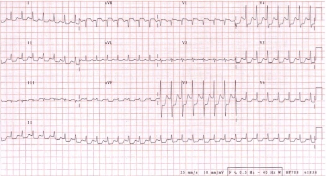
ECG 1
Using the ECG in 20 Seconds Method so we don’t miss anything:
Rate: 29 x 6 = 174
Rhythm: There are no P waves to be see, but the QRS are regular.
QRS: Narrow complexes- not too tall, not too small, normal morphology.
Axis is normal
ST-T: There is global ST depression in all leads, with ST elevation in aVR and V1
PR/QT: QT appears prolonged.
Conclusion: This is a narrow complex supra ventricular tachycardia with ischaemic ST changes.
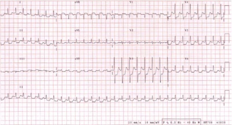
ECG 2
Rate: 17 x 6 = 102
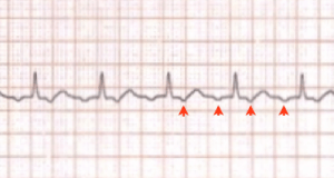
Rhythm: There are P waves. Are up in II and inverted in aVR? NO- they are the opposite to this. It may indicate limb lead reversal, but if we look at I, the pave is up as it should be. The other diagnosis then is atrioventricular ectopics, i.e. the p wave is generated in the AVN but by ectopic focus. The P waves are blocked and we see in lead II that there are 2 paves to every QRS, so it is an ectopic focus with a 2:1 block. Look at lead II below.
QRS: Normal morphology
Axis: Normal
ST-T: The ischaemic changes have resolved, although a minor amount of ST depression in V2
PR/QT: The QT interval appears still slightly prolonged. Let’s calculate the QTc by using Bazetts’ formula
QTc = QT/√RR
QTc = (8×0.04)/√(16×0.04) = 0.32/√.64 =0.32/0.8 =0.4, so it is not prolonged.
Conclusion: The second ECG has seen the almost total resolution of ischaemic changes, but there is an ectopic atrial rhythm with a 2:1 block.


