

The Lewis lead(1), is a means of detecting atrial activity and can be used where p waves are not readily evident. It is particularly helpful in wide complex tachycardias, where the presence of atrial activity can be used to exclude a diagnosis of ventricular tachycardia. It is meant to be seen on the cardiac monitor and not the ECG.
How to do it
We will be monitoring lead I. The electrodes need to be placed in the following positions: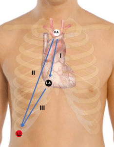
-The right arm lead is placed over the manubrium
-The left arm lead is placed at the right sternal border of the 5th intercostal space and
-The left leg lead is placed over the right costal margin
An example from the Journal of emergency Medicine (2) illustrates this well.
A 52 year old woman presents with dyspnoea and shock. The patient is diagnosed with a pneumonia and treated. The 12 lead ECG is shown below. Ventricular Tachycardia is considered as the diagnosis.
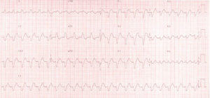
The Journal of Emerg Med August 2012
A Lewis Lead placement was performed, with increased amplitude and showed P waves, allowing the diagnosis of ventricular tachycardia to be excluded.
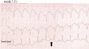
The Lewis lead ECG
The Lewis lead has always been meant to be used on a cardiac monitor. However a modified Lewis Lead ECG has been proposed(3).
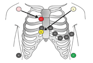
The right arm electrode is moved to the second intercostal space parasternal right and the left arm electrode to the fourth intercostal space parasternal right.
In this study there was a much improved detection of ventriculoatrial conduction in the lewis ECG.
Other Links
Diagnosing Ventricular tachycardia in 5 easy steps
References
- Lewis,T. Auricular Fibrillation. Clinical Electrocardiography 1913. pp86-97
- Rodrigues de Holanda-Miranda et al.Lewis lead enhances atrial activity detection in wide QRS tachycardia. The J oe Emerg Med. August 2012, Vol 43,Issue 2; ppe97-e99
- Huemer M et al. The Lewis lead for detection of ventriculoatrial conduction type. Clin Cardiol. 2016:39,2; pp126-131


