
Here are my top 8 tips for succeeding at intubation.
1. Use a Video Laryngoscope(VL), or at least have one nearby
Okay, maybe a little controversial still. But if there is some way that you can get your hands on one, have it on standby. In a statement several years ago, Ron Walls said that VL was the standard of care and we should be using it. There was an outcry! People were concerned: “We need to know how to use a normal laryngoscope, what if the video laryngoscope doesn’t work, or I go to an ED without one?”
I understand, I hear you. But……..if you can use a normal laryngoscope, why not use a VL such as a C-MAC( I have no financial interest to declare) or similar, where the blade is the same and you can use it as a normal laryngoscope or kick it into gear and get the help you need.
Personally I always feel better when I know I have one there. How often might I need to use it? Less than 5% of the time.
2. Time: Make sure I have the maximum amount of apnoea time available to me
I am and have always been a massive fan of apnoeic oxygenation. I do whatever I can to maximise oxygen reserves in my patients. I use the nasal prongs at high flow, I use CPAP before intubation, I use sitting up and delayed sequence intubation…..whatever I can to buy me time.
3. Position: Tragus of the ear to Sternal Notch
We’ve covered this so many times, but it’s important to do so once again. The aim is to lift the head so that the ear and the sternal notch are in line. This brings the cords into view. Obviously not in the suspected cervical injury. That’s where VL comes into use.
4. Sweep the blade to the left and hold in the midline
When inserting the laryngoscope blade, do so to your right(ie., the right side of the patient’s mouth) and sweep the tongue towards the left, BUT let the blade come to rest in the midline. Why? Because you want to give yourself maximum room to the right of the patient’s mouth to be able to see where you are inserting the tube giving you the best view of the anatomy(the midline view). If it helps get a friend to pull the patient’s right cheek back, so you can get a better view.
5. Perform Epiglotoscopy
The most important landmark when looking for the cords are not the cords, but the epiglottis. Find the epiglottis and you will find the cords. The best way to do this is to find the back of the tongue and then move slowly downwards. The single biggest mistake I see, is that the blade is introduced in too far to begin with. It’s past the epiglottis. If this happens, just pull the blade back and the epiglottis will flop into view.
6. Don’t pivot the blade unless you want to break the teeth
In patients with dentures, we leave those dentures in whilst bagging, as they scaffold the mouth, so we can ventilate. When it’s time to intubate we remove them. In patients who have their own teeth, one of the risks we run is to break those teeth. Do that and you’re in a world of hurt, as you now have broken teeth that need repair, but you may also have fragments that will need to be retrieved via bronchoscopy at some point. The technique is not swivelling or pivoting the laryngoscope, but pulling out in the direction of the handle.
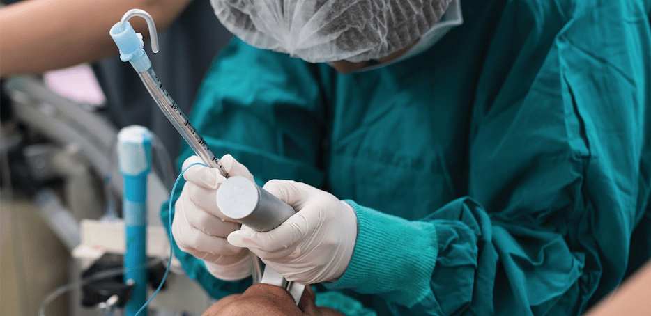
7. Always use a stylet
I always use a stylet. There is the argument of bougie vs stylet. My argument is that the angle at the end of the stylet and the bougie is the same. If there is any swelling of the airway, the stylet may be better. Also if it is a very anterior airway, the angle of the stylet can be manipulated to what you need.
Irrespective of which you use. Use one.
8. Only rely on End Tidal CO2(ETCO2) for verification of tube placement
“I saw it go through the cords” may be famous last words. I’ve said them myself. I saw the tube go through, but the sats are dropping and there is no ETCO2 trace………the tube’s not in. Pull it out and do it again.
Misting of the tube is unreliable. Auscultating for breath sounds is misleading, as a tube in the oesophagus may generate sounds that appear to be coming from the lungs.
ETCO2 is the gold standard. Wait for several waveforms. Read the Blog on Capnography.


