A 50 male patient is brought to your emergency department by ambulance post cardiac arrest. He has an adrenaline infusion running at 100mcg/min, has been intubated in the field by paramedics and has had a protracted resuscitation period with 10mg of adrenaline given in the field, for repetitive loss of ROSC.
The patient is very unstable and you are losing ROSC regularly, plus the is repeatedly going into Ventricular Fibrillation. You are overseeing the almost continuous CPR and notice blood coming from the endotracheal tube. Now this blog is not about treating arrhythmias, but about what happens next, when the lab calls.
To read more about Electrical Storm and Double Sequential Defibrillation, read here. You can watch my lecture on The Resistant Arrhythmia below.
The nurse hands you the phone and says..
“It’s the lab with a critical result.”
“Really? Every result is going to be critical on this patient!”
But this result really is. The patient’s blood isn’t clotting; they’re in Disseminated intravascular Coagulation (DIC).
What is DIC?
The International Society on Thrombosis and Haemostasis gives the following definition:
“An acquired syndrome characterized by the intravascular activation of coagulation with loss of localization arising from different causes. It can originate from and cause damage to the microvasculature, which if sufficiently severe, can produce organ dysfunction.”(1)
It’s the body’s activation of systemic coagulation and systemic inflammation, always secondary to some other condition ie., It’s not a primary disorder, it is a response. This systemic response results in two things:
- Deposition of Fibrin causing microvascular thrombi leading to multi-organ failure
- Clotting factor and platelet consumption leading to haemorrhage
Causes of Acute DIC:
- Infective: Sepsis and Septic shock caused by bacteria are by far the most common causes, however viruses and Fungi or Parasites can also cause it.
- It can occur in up to 50% of patients with sepsis(2)
- It is an independent predictor of mortality and significantly increases mortality(3)
- Traumatic: Neurotrauma is a specific cause
- Malignancy: Haematologic malignancies as well as metastatic malignancy
- Transfusion Reactions: Transfusion and Haemolytic Reactions
- Obstetric Causes: Amniotic Fluid Embolism, Abruptio Placentae, Preeclampsia/eclampsia, HELLP syndrome, Septic Abortion, Retained dead fetus,
- Hepatic Failure
- Toxinology: Envenomation
- Environmental: Hyperthermia, Heat stroke
CLINICAL
DIC is a consequence of the underlying condition. Remove the condition and the DIC may improve. That may be possible where we can quickly correct the underlying disorder. For example, a patient with significant sepsis or metastatic malignancy does not have something that is quickly reversible. However the eclamptic patient, or that patient with abruptio placentae, may be amenable to more rapid treatment and reversal.
Clues to the presence of DIC include:
- Bleeding (the most common finding)
- From mucous membranes or GIT
- Petechiae and ecchymosis
- Renal and hepatic failure
- Shock
- Respiratory failure
Making the Diagnosis
Diagnosing DIC clinically, can be very challenging. For example, differentiating between liver failure induced coagulopathy and DIC can be difficult.
Laboratory Tests rely on:
- aPTT/PT times
- Usually prolonged, although may be normal
- D-dimer
- Platelet count
- Significant thrombocytopaenia is present in most patients
- Clotting factors/inhibitors
Scoring Systems can help.
THE DIC DIAGNOSTIC ALGORITHM(1):
To be used, the patient must have a disorder known to be associated with DIC.
The following coagulation tests need to be ordered:
-
- Platelet Count
- Prothrombin Time
- Fibrinogen
- Soluble Fibrin Monomers or
- Fibrin Degradation Products(FDP)
- We can then assign scores to the test results
- Platelet Count
- >100 = 0 points,
- <100=1 point
- <50=2 points
- Platelet Count
- Elevated FDP
- no increase =0 points
- moderate increase=2 points
- strong increase =3 points
- Prolonged Prothrombin time
- <3sec=0points,
- >3sec but <6sec =1point,
- >6 sec=2 points
- Fibrinogen level
- >1.0g/L = 0 points
- <1.0g/L=1 point
The DIC Score Calculator has been prospectively validated with a sensitivity as high as 93%. A score of > 5 indicates DIC.
MANAGEMENT OF ACUTE DIC
Treat the underlying condition
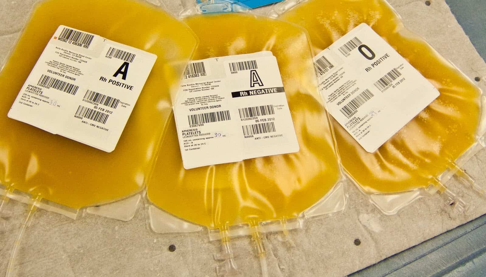
If sepsis give antibiotics and replacement fluids etc In terms of replacement products, you MUST consult with your Haematologist as to what to give and doses. Below is a rapid review of what the lab will provide you with.
Fresh Frozen Plasma(FFP)
- In bleeding and abnormal coagulation
- It must be ABO compatible.
- 4 units (15ml/kg rapidly infused
FFP contains all the coagulation factors except platelets
- Fibrinogen
- Albumin
- Protein C
- Protein S
- Antithrombin
- Tissue Factor Pathway Inhibitor
It does not contain erythrocytes or leukocytes
Cryoprecipitate
- Contains Fibrinogen
- Aim to keep Fibrinogen levels above 1g/L
- Usually give 1 pool, which is 5 bags in an adult patient. Each bag provides 325mG of Fibrinogen.
- In children it is usually 1 unit per 10kg wight of the child.
Cryoprecipitate contain:
- Fibrinogen
- Von Willebrand factor
- Factor VIII
- Factor XIII
- Fibronectin
Platelets
- In bleeding patients transfuse platelets when platelet count drops 50 x 109/L
- In those patients not bleeding, transfuse when platelets drop below 20 x 109/L
References
- Taylor FB Jr, Toh CH, Hoots WK, Wada H, Levi M. Towards definition, clinical and laboratory criteria, and a scoring system for disseminated intravascular coagulation. Thromb Haemost. 2001 Nov. 86(5):1327-30.
- Levi M, Toh CH, Thachil J, Watson HG. Guidelines for the diagnosis and management of disseminated intravascular coagulation. British Committee for Standards in Haematology. Br J Haematol. 2009 Apr. 145(1):24-33.
- Levi M,M et al. Disseminated Intravascular Coagulation. Oct 7 2018, https://emedicine.medscape.com/article/


