
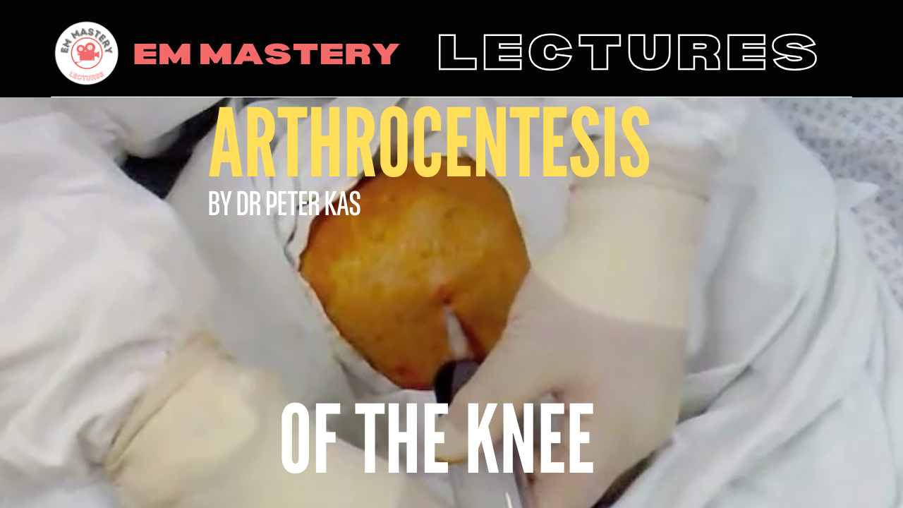
A 70 yo patient presents to the emergency department with a painful swollen knee. There has been no trauma and he is systemically well. The decision is made to perform arthrocentesis of the knee joint.
The Procedure:
- I prefer to use the parapatellar approach. Other approaches are available, however I explain why I use this approach in the video below.
- The following is performed with a sterile technique.
- Prepare the skin with sterile solution, allow to dry.
- Drape the area
- Apply local anaesthetic to the skin in the area where the needle is to be inserted into the knee joint.
- Take a 60 mL syringe, although a 20mL syringe will work as well. Attach an 14G or 18G needle.
- Identify the landmarks:
- Locate the midpoint of either the medial(my preferred technique, the reason explained in the video below) or the lateral border of the patella.
- Insert needle below the midpoint in a direction perpendicular to the skin pointing towards the intercondylar notch of the femur. A rolled towel under the knee can result in a slight flexion of the joint, which may facilitate needle entry into the joint.
- Aspirate until synovial fluid is extracted. This usually occurs at about 1-2 cm.
- Aim to not walk the needle along the bone. If bone is encountered, withdraw the needle and gently reinsert.
- Once the procedure is complete, place a sterile bandage over the area.
View the video of the procedure below.
This is content for subscribers.
For less than the price of a cup of coffee a week, you can get full access to everything
Published on: 30/01/2025