
A 19yo presents with a complaint of palpitations lasting several seconds and some chest tightness that lasts a few minutes, but is gone when the patient retracts his arms posteriorly to stretch his chest.
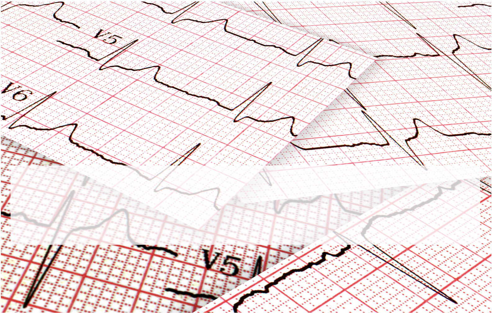
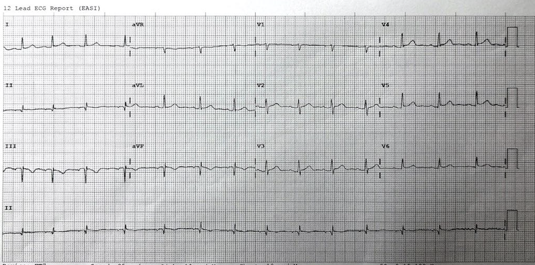
His ECG shows inverted T wave inferiorly. A high sensitivity troponin is sent and comes back normal. The ECG is shown below.
What does the ECG show?
(a) Acute Inferior Infarction
(b) Old Inferior infarction
(c) Pericarditis
(d) None of the above
What would you do next?
Answer
This is an ECG we have covered before in one form or another.
The answer is (d)
When looking at the ecg and using the ‘ECG in 20 Seconds’ approach it shows:
Rate:84 bpm
Rhythm: Sinus rhythm
P axis: the p wave is upright in aVR and inverted in lead II- therefore this is LIMB LEAD REVERSAL. In a normal P wave axis ie., sinus rhythm, the p wave must be upright in II and inverted in aVR.
When the leads are correctly placed the following ECG was obtained:
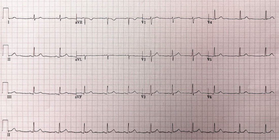
Troponin was normal. It was repeated and still normal.
The Heart Score was low indicating a low risk patient.


