
He is haemodynamically stable, with a normal clinical examination.
His ECG is shown below.
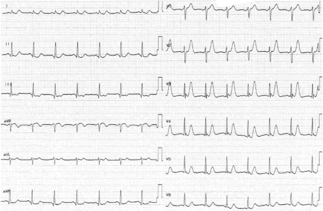
There are definitely hyper acute T waves. What does that actually mean? It’s important to note that here is no formal definition of a hyper acute T wave. However it is not based on amplitude alone.
The hyper acute T wave is considered a STEMI-equivalent.
Koechlin L et al(1) conducted a post hoc analysis of a multicenter diagnostic study and looked at the diagnostic performance of T wave amplitude for diagnosing myocardial infarction. They found that they were not useful in making this diagnosis.
One of the concerns is that they only looked at T wave amplitude. Smith(2) states that more than just amplitude is important to make the diagnosis. He refers to the T-wave ‘bulk’ The T wave is large in area, symmetric relative to the QRS.
The term ” T wave towers” over the R wave in V3, or even that the QRS can fit into the T wave are important in terms of this ‘bulk’.
References
- Koechlin L et al. Hyperacute T Wave in the Early Diagnosis of Acute Myocardial Infarction. Ann of Emerg Med.2022; 1-9
- Smit S et al. Hyperacute T-waves Can be a Useful Sign of Occlusion Myocardial Infarction if Appropriately Defined. Annals of Emerg Med. 2023;1-4


