
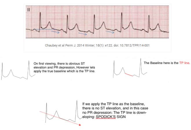
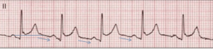
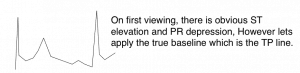
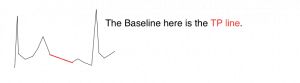
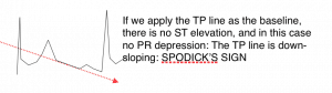
Spodick’s Sign appears in Stage I of Pericarditis and is a downsloping of the TP line ie., the baseline. It is said to be present in some 80% of cases of acute pericarditis and is best visualised in lead II and the lateral precordial leads(1).
Chaubey et al Perm J. 2014 Winter; 18(1): e122. doi: 10.7812/TPP/14-001
The ECG findings of pericarditis are important to know as they are present in about 90% of cases(2).
Stages of Pericarditis
Stage 1
Diffuse ST elevation in all except V1 and aVR and PR depression
Stage 2
Normalisation of segments
Stage 3
T-wave inversions
Stage
Normalisation of the ECG(3)
These ECG abnormalities result from inflammation of the subepicardial myocardium resulting in the resulting PR segment depression(4).
To learn more about Pericarditis, read the blog on this topic.
How good is Spodick’s Sign?
It seems to be accepted as a reliable sign, although there are no controlled studies looking at it. I believe one is under way at present. We shall see.
I use it.
References
- Chaubey VK et al. Spodick’s Sign: A Helpful Electrocardiographic Clue to the Diagnosis of Acute Pericarditis. Perm J. 2014 Winter; 18(1): e122. doi: 10.7812/TPP/14-001
- Spodick DH. Diagnostic electrocardiographic sequences in acute peri- carditis: significance of PR segment and PR vector changes. Circula- tion. 1973;48:575-580
- Spodick DH. Acute pericarditis: current concepts and practice. JAMA 2003;289:1150.
- Makaryus J et al Spodick’s Sign. August 2008, Volume 121, Issue 8, Pages 693–694
Other References
Chabra L, Spodick DH. Ideal isoelectric reference segment in pericarditis: a suggested approach to a commonly prevailing clinical misconception. Cardiology. 2012;122(4):210–2. DOI: http://dx.doi.org/10.1159%2F000339758
Masek KP, Levis JT. ECG diagnosis: acute pericarditis. Perm J. 2013 Fall;17(4):e146. DOI: http://dx.doi.org/10.7812/TPP/13-044


