
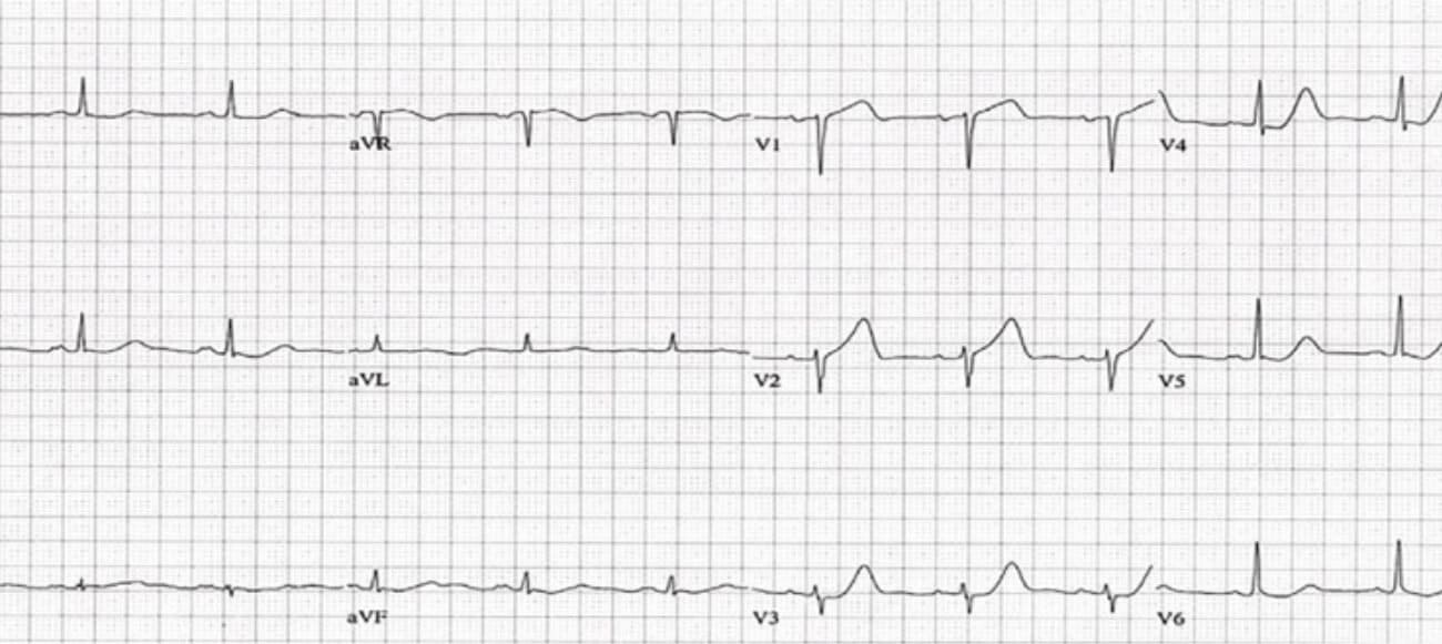
A 65 year old woman presents with a 20 minute episode of chest pain. Her ECG is shown below. Are there any concerning features for ischaemia?
Source: The west journal of EM 18(4):601-606 · June 2017(1)
Significance of an upright T wave in V1
Normally the T wave is inverted or flat in lead V1. An upright T wave in V1 may be considered a normal variant. However assuming that there is no ventricular hypertrophy or LBBB, a large, upright T wave in V1 may be abnormal if:
- The T wave in V1 is taller than the T wave in V6. (This is referred to as a loss in precordial T wave balance(2).)
- The upright T wave in V1 is new.
Patients with upright T waves in V1 are considered to be 4 times more likely to have significant coronary artery disease than those without(3).
Causes of upright T waves in V1
- Ischaemia: This can be single or multiple vessel disease and involves one or more of the following:
- Left Circumflex
- Right Coronary Artery
- Left Anterior Descending
- Left Ventricular Hypertrophy
- LBBB
- High Left ventricular voltage
- Lead misplacement
Keep on the lookout for T wave changes in the precordial leads. This is one of those subtle changes to be aware of.
References:
- Tewelde S et al. Pitfalls in Electrocardiographic Diagnosis of Acute Coronary Syndrome in Low-Risk Chest Pain. The Western Journal of Emergency Medicine 18(4):601-606 · June 2017
- Pinto J J et al. Tall upright T waves in the precordial leads. Circulation 1987;36(5):708-16
- Manno B.V et al. Significance of the upright T wave in precordial lead V1in adults with coronary artery disease. JACC Volume 1, Issue 5, May 1983
Peter Kas


